Casemasters Blog
Microscopic Dentistry
A recent article entitles “The Effect of the Dental Operating Microscope on the Outcome of Nonsurgical Root Canal Treatment: A Retrospective Case-control Study” investigated the effect of using dental microscopes on the success rate of endodontic treatment of maxillary first molars. This article included 190 molar teeth that required endodontic retreatment. These molar teeth were divided into 2 groups: first group had their initial RCT using a microscope and the second group without a microscope. MB root was 3 times more likely to develop periapical lesion when the initial treatment was performed without microscope. The conclusion of this article was that operating microscopes can aid in proper education and can help dentists to achieve good knowledge and insights into cases that need advanced training and modern visualization tools such as microscope.
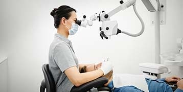
Dental microscopes are important for magnification to enhance precision, quality and longevity of treatment. Dental procedures should be minimally invasive with low risk and short operative/healing time. Proper visualization is important for quality dental work and dental loupes/microscope are increasingly becoming essential dental armamentarium.
Dental microscopes are also becoming essential in dental education and have been introduced in some dental academic curricula, especially in light of the availability of additional attachments such as camera and video devices, which allow dental student to engage with their educators and to see the methodology and the results of dental procedures. In addition, in dental practices, patients who get treated with the aid of microscopy can feel more involved in treatment and better understanding the treatment process.

The modern practice of dentistry such as veneers, soft-tissue management and even restoratives require clear vision and appropriate magnification. There are currently different commercially available dental microscopes in the market and a dentist should consider certain criteria before deciding to purchase a dental microscope. Major manufacturers of dental microscopes include but not limited to AmScope, Global Surgical Corporation, Leica and Zeiss.
The microscope head can be binocular to allow the dentist to get as realistic view as possible. This microscope has adjustable eyepieces so that magnification can be changed according to the dental procedure to be done. Other microscopes can allow for adding another head (teaching head) which enable 2-3 people to work together in one operation. These microscopes have independent pivot and separately adjustable magnification levels that fit with each member of the dental teamwork.
Optical quality (resolution/depth of field/light transmission) and illumination are other important features of dental microscope. High resolution improves the clarity of images, large depth of field provides better view of the area of interest and maximum light transmission provides good visibility. Illumination contributes to brightness and color which is determined through different types of light sources (Halogen/LED) which, in turn, have different life spans and light/heat intensities.

The functionality and design of the microscope are also important. A dental microscope should be easy to work with, easily mounted and integrated in the practice environment, lightweight and stable in order to ensure seamless workflow. Ergonomic design of the microscope should consider patients’ comfort and avoid dentist’s repeated movements and unhealthy working position that could have serious consequences. The design of the microscope should also adapt with the body frame of the dentist so that he/she need not to strain to get to the right position to tend to the patient. The dental microscope should be easy to clean with antimicrobial and to maintain.
A percentage of 7% of restorative treatment nowadays is done as replacement of existing fillings and such cases clearly require using dental microscopes with high efficiency. Using microscopes in such cases increases the quality of dental work, productivity of dentist and patients’ education.