Casemasters Blog
Ready to Use Titanium-Based Nanostructures for Dental Implants
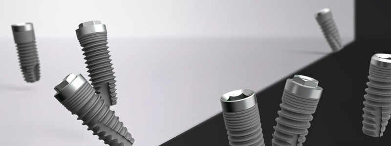
Titanium alloys have excellent mechanical properties, corrosion resistance and biocompatibility, but their usage as dental implants is compromised by bacterial growth causing infection, inflammation and peri-implantitis. The infection process starts with the formation of bacterial biofilms, that is, bacteria accumulate over the implant’s surfaces and form a matrix of extracellular polymeric substances to protect themselves from host immune system and antimicrobial agents. The commonly followed strategy for treating peri-implantits is by surgical removal of the implant, tissue debridement and then reimplantation after average 6 weeks.
This recovery process accounts for 2 million cases of failure each year and $5 billion loss only in USA. Dental implants’ manufacturers work currently on releasing new products with antimicrobial coatings which can be antibiotics, oxidants or biocides to kill bacteria and prevent biofilm formation. However, long-term results are not encouraging as the concentration of antimicrobial agents depletes and antimicrobial resistance can develop.

The early results of nanostructured dental materials are encouraging as they show good effect in inhibiting bacterial growth and adhesion with their surfaces. Titanium-based nanostructures could delay biofilm formation for at least 6 days by means of mechanical rupture of bacterial cell membranes. These nanoparticles have self-cleaning effect that can sweep away microorganisms and inhibit adhesion of Staphylococcus aureus. They can pierce bacterial cell membranes, interfere with DNA synthesis and cause bacterial inactivity or death. Titanium nanoparticles synthesis can be completed through hydrothermal methods which enhance osteo-integration and prevent biofilm formation. The interaction of bacterial cells and human body cells on one hand and the implant’s surface on the other hand is visualized through several devices, such as confocal Laser scanning microscope (CLSM) and focused-ion-beam scanning electron microscope (FIB-SEM).