Casemasters Blog
Surgical Simulation of Cyst Removal: Never Easier
Fenestration of the maxillary sinus is considered the major complication of the surgical removal of maxillary cysts. Therefore, many oral surgeons currently tend to apply piezosurgery for cyst removal as it is capable of preserving the structure of the sinus through its modulated frequency that permits safe, accurate bone cutting without harming the surrounding soft tissues. 3D scanning/modeling can also aid in diagnosis, preoperative planning and even simulation of surgery to reduce complications and surgical trauma.
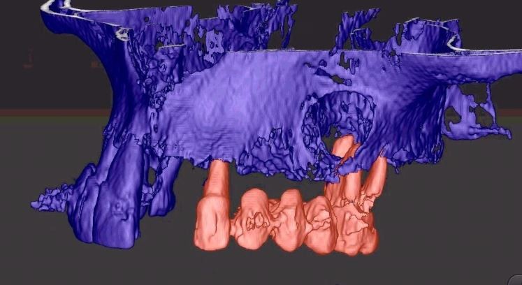
3D modeling was first applied with plaster or resin objects mimicking the maxillofacial morphology; the creation of such object was based on 3D image constructions where certain structures such as cyst, nerves or blood vessels are colored to accurately visualize their shape, size, position and course. With the advances in 3D printing technology, it is now possible to create models with hybrid materials, each with specific density and color similar to bone, teeth or cartilage. This can offer a realistic simulation of surgery with appropriate hardness and accurate representation of hard tissue deformities. Osteotomy can be practiced on these models while the teeth and their roots as well as vital structures like alveolar nerve and maxillary sinus are all observed. These 3D-printed models have been tested for almost every oral and maxillofacial procedures including mandibular reconstruction surgery, extraction of impacted molars, periodontal tissue regeneration and cyst removal.
In literature, there are several surgical approaches reported for the maxillary area such as Caldwell-Luc surgery and endoscopic sinus surgery. Choosing the suitable surgical approach depends on the existence of sufficient surgical field, the amount/extension of bone defects and presence/absence of impacted tooth. Tissue scarring as well as intracavernous/sinus-wall penetration is among the most common complications that might arise in case of insufficient preoperative planning.

3D-printed models can thus clearly identify each structure and guide the surgeon on each bone fragment to be removed or preserved. In addition, with piezoelectric surgical device a surgeon can manage for minimal bone cutting, least soft-tissue trauma and even bone grafting while avoiding surgical complications such as prolonged oedema and paresthesia after surgery.